
Description
PrymaLab · Research Use Only
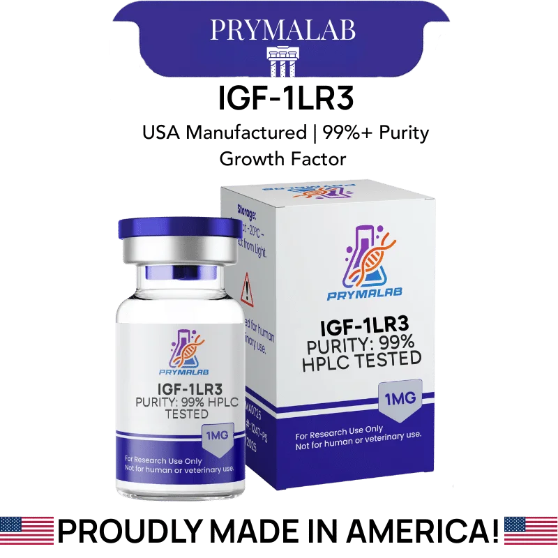
IGF1 LR3 1MG
Long R3 IGF-1 · 83 residues · CAS 946870-92-4 · ~100-fold reduced IGFBP affinity
IGF-1 LR3 1mg supplies Long R3 insulin-like growth factor 1, an 83-residue analog with molecular weight 9117.60 and CAS number 946870-92-4. Two modifications distinguish it from the native molecule: an arginine substitution replacing glutamate at position 3, and a 13-residue extension at the amino terminus.
Specification Table
| Property | Value |
|---|---|
| Compound | Long R3 IGF-1 |
| Alternative names | IGF-1 LR3, IGF1 LR3, LR3 IGF-1 |
| CAS number | 946870-92-4 |
| Molecular formula | C400H625N111O115S9 |
| Molecular weight | 9117.60 g/mol |
| Residue count | 83 amino acids |
| Parent molecule | Human IGF-1 (70 residues) |
| Modification 1 | Arginine substituted for glutamate at position 3 |
| Modification 2 | 13-residue N-terminal extension, sequence MFPAMPLLSLFVN |
| Primary molecular target | Type 1 IGF receptor (IGF-1R) |
| Binding protein affinity | Reduced approximately 100-fold across IGFBP-1 through IGFBP-6 |
| Reported half-life | Substantially extended relative to native IGF-1 |
| Disulfide bonds | Three, conserved from the parent molecule |
| Production route | Recombinant expression |
| Vial content | 1 mg lyophilized powder |
| Appearance | White lyophilized cake |
| Purity | Per lot-specific certificate of analysis |
| Solubility | Soluble in dilute acetic acid and in bacteriostatic water |
| Storage, lyophilized | -20°C, protected from light and moisture |
| Storage, reconstituted | 2-8°C, protected from light |
| Reconstituted stability | Not established under arbitrary conditions |
| Regulatory status | No approved human or veterinary formulation in any jurisdiction |
What Do the Two Modifications Do?
The name encodes the chemistry. Long refers to the 13-residue N-terminal extension, and R3 to the arginine substituted in at position 3.
Native IGF-1 carries glutamate at position 3. Substituting arginine changes the local charge at a site that participates in binding-protein recognition, and the substitution alone measurably reduces IGFBP affinity. The 13-residue extension, sequence MFPAMPLLSLFVN, adds further steric interference at the same interface while also originating from the recombinant expression construct.
Together the two changes reduce affinity for the full binding-protein family, IGFBP-1 through IGFBP-6, by roughly a hundredfold. Receptor binding at IGF-1R is retained.
The design logic parallels des(1-3)IGF-1 but arrives by a different route. One deletes the interaction site. The other substitutes and extends around it. Both leave the receptor-binding face untouched.
Why Does Binding Protein Evasion Change the Pharmacology?
Roughly 97 percent of circulating IGF-1 is not free. It sits in ternary complexes with IGFBP-3 and the acid-labile subunit, a reservoir arrangement that buffers free growth factor concentration and extends the pool half-life while keeping receptor-available ligand tightly controlled.
That reservoir is why native IGF-1 has a short free half-life measured in minutes despite persisting in plasma for far longer in bound form. The binding proteins are doing two jobs at once: protecting the molecule from clearance and withholding it from receptors.
An analog that escapes capture inverts the arrangement. More of the administered material stays free and receptor-available, and clearance behaviour changes accordingly. Reported half-life for IGF-1 LR3 is substantially longer than for native IGF-1, and the practical consequence is that a given amount produces both greater and more prolonged receptor engagement.
For experimental design that means exposure duration becomes a variable that has to be managed deliberately rather than assumed to track the exposure schedule.
How Should IGF-1 LR3 1mg Be Normalised Against Other IGF Analogs?
By moles, always. The numbers make the reason obvious.
Native IGF-1 is 7649 daltons across 70 residues. des(1-3)IGF-1 is 7371.48 across 67 residues. IGF-1 LR3 is 9117.60 across 83. Comparing equal masses of LR3 and des(1-3)IGF-1 delivers roughly 24 percent fewer molecules of the former, before any difference in binding-protein behaviour or clearance is considered.
One milligram of IGF-1 LR3 is approximately 110 nanomoles. The same mass of des(1-3)IGF-1 is approximately 136 nanomoles. Any comparative design working from milligram equivalence starts with a systematic bias of that size baked in.
Add the differing half-lives on top and mass-based comparison becomes untenable. Molar normalisation plus an explicit statement of exposure window is the minimum for a comparison that means anything.
What Are the Practical Limits of This Compound?
Recombinant protein identity is the first issue worth raising. IGF-1 LR3 is expressed in a host organism rather than synthesised stepwise, which means correct disulfide pairing and correct folding are manufacturing findings rather than guarantees. Three disulfide bonds have multiple possible pairings, and a misfolded preparation registers as protein by mass while lacking activity. Mass alone does not establish that a preparation is functional.
Insulin receptor cross-reactivity applies here as it does across the IGF family. Structural homology means that at sufficient concentration the analog engages the insulin receptor, and a metabolic readout without a discriminating control does not establish which receptor produced it.
Finally, the reduced binding-protein affinity that makes the analog useful also removes a natural buffering mechanism. In systems where IGFBPs normally moderate free ligand concentration, that regulation is absent, and concentration control has to come from the experimental design instead.
How Is Recombinant Identity and Folding Confirmed?
This compound is expressed in a host organism rather than assembled stepwise, which introduces failure modes that synthetic peptides do not have. Correct disulfide pairing is a manufacturing finding, not a design guarantee.
Mass spectrometry confirms molecular weight. With peptide mapping after enzymatic digestion it confirms sequence as well. What it does not readily confirm is disulfide connectivity, since three bonds can pair in several arrangements that all give the same total mass. Distinguishing them requires digestion under non-reducing conditions followed by careful fragment analysis, which not every certificate of analysis reflects.
Reversed-phase chromatography separates correctly folded material from misfolded variants, because folding state changes surface hydrophobicity and therefore retention. A single sharp peak indicates conformational homogeneity. Shoulders or multiple peaks indicate a mixture, and a preparation that is 95 percent pure by mass may still be a mixture of conformers.
Size-exclusion chromatography catches the other common problem, which is aggregation. Growth factor proteins aggregate under freeze-thaw and mechanical stress, and aggregated material is both inactive and capable of interfering with the assay it enters. Running it costs little and blocks a class of uninterpretable experiments.
What the Extended Half-Life Costs
Longer persistence is usually sold as an unqualified advantage. In experimental work it carries costs, and they are worth stating plainly.
Receptor downregulation is the first. Sustained agonist occupancy drives internalisation and degradation of many receptor tyrosine kinases, so prolonged exposure can reduce the very signalling it was intended to produce. A short exposure and a long one at the same concentration are not comparable conditions, and a declining response over time may reflect receptor loss rather than any change in the ligand.
Washout is the second cost. An analog that persists is difficult to remove from a system, which complicates any design needing a defined exposure window followed by a clean recovery period. Medium changes remove free ligand but not what has already been internalised.
Third, extended exposure narrows the interpretive value of endpoint measurements. When a compound is present throughout, a measurement at the end integrates everything that happened, and separating early signalling from late adaptation requires a time course that many designs omit.
Reconstitution and Storage in Laboratory Practice
One milligram is a small amount of material spread across a large molecule, so reconstitution volume drives the entire downstream concentration range. Decide the target working concentration before opening the vial rather than after.
Dilute acetic acid is the conventional reconstitution medium, since this protein class is more soluble and more stable at mildly acidic pH. Add diluent slowly against the vial wall and allow dissolution without agitation. Vortexing introduces shear that can disrupt the folded structure, and a disrupted preparation will not announce itself visually.
Carrier protein reduces adsorptive loss at low working concentrations, which is a genuine and often underestimated source of variability with recombinant growth factors. Aliquot before freezing to avoid repeated freeze-thaw. Hold lyophilized material at -20°C. Record lot, diluent, reconstitution volume, resulting concentration and date for every vial opened.
Published Literature
Each reference below was confirmed against the publisher record or a primary index. Baxter is included for the ternary complex biology rather than for the analog itself.
- Francis GL, Ross M, Ballard FJ, Milner SJ, Senn C, McNeil KA, Wallace JC, King R, Wells JR. Journal of Molecular Endocrinology. 1992;8(3):213-223.
- Tomas FM, Knowles SE, Chandler CS, Francis GL, Owens PC, Ballard FJ. Journal of Endocrinology. 1993;137(3):413-421.
- Bagley CJ, May BL, Szabo L, McNamara PJ, Ross M, Francis GL, Ballard FJ, Wallace JC. Biochemical Journal. 1989;259(3):665-671.
- Baxter RC. American Journal of Physiology: Endocrinology and Metabolism. 2000;278(6):E967-E976.
Frequently Asked Questions
What is IGF-1 LR3 1mg?
IGF-1 LR3 1mg is a lyophilized vial of Long R3 insulin-like growth factor 1, an 83-residue analog with CAS number 946870-92-4 and molecular weight 9117.60. It carries an arginine substitution at position 3 and a 13-residue N-terminal extension. Supplied for laboratory research use only.
What does the name Long R3 mean?
Long refers to the 13-residue N-terminal extension, sequence MFPAMPLLSLFVN. R3 refers to the arginine substituted for glutamate at position 3 of the native sequence. Both modifications interfere with binding-protein recognition while leaving the receptor-binding face of the molecule intact.
How much does IGFBP affinity change?
Roughly a hundredfold reduction across the full binding-protein family, IGFBP-1 through IGFBP-6. Since about 97 percent of circulating native IGF-1 is sequestered in ternary complexes with IGFBP-3 and the acid-labile subunit, escaping that capture substantially changes how much of the molecule remains receptor-available.
How does IGF-1 LR3 differ from IGF-1 DES?
IGF-1 LR3 is 83 residues at 9117.60 daltons and achieves binding-protein evasion through substitution and extension, with a substantially extended half-life. IGF-1 DES is 67 residues at 7371.48 daltons and achieves it by deleting the N-terminal Gly-Pro-Glu tripeptide, with faster clearance. They suit different experimental windows.
Why must comparisons be made by moles rather than mass?
One milligram of IGF-1 LR3 is approximately 110 nanomoles. The same mass of des(1-3)IGF-1 is approximately 136 nanomoles, roughly 24 percent more molecules. Any comparative design working from milligram equivalence carries that systematic bias before differences in clearance or binding-protein behaviour are even considered.
Does molecular weight confirm the protein is functional?
No. IGF-1 LR3 is a recombinant protein with three disulfide bonds, and those bonds have multiple possible pairings. A misfolded preparation registers correctly by mass measurement while lacking receptor binding entirely. Functional confirmation requires a binding or activity assay, not a mass check.
What controls does IGF-1 LR3 work require?
One control separating IGF-1 receptor signalling from insulin receptor signalling, because the two receptors are structurally related and cross-engagement happens at sufficient concentration. Beyond that, remember that reduced binding-protein affinity strips out a natural buffering mechanism, so free ligand concentration now has to be controlled by the design instead.
Why use dilute acetic acid for reconstitution?
This protein class is more soluble and more stable at mildly acidic pH than at neutral pH. Add diluent slowly down the vial wall and allow dissolution without agitation. Vortexing introduces shear that can disrupt the folded structure, and structural disruption is not visible in the reconstituted solution.
How should the vial be stored?
Dry cake at minus 20 Celsius, sealed against light and moisture. Reconstituted solution between 2 and 8 Celsius in the dark, aliquoted before the first freeze. Carrier protein in the diluent is worth considering, since adsorptive loss at low working concentrations is larger than most people expect.
How is correct folding confirmed?
Reversed-phase chromatography separates correctly folded material from misfolded variants, since folding state changes surface hydrophobicity and therefore retention. A single sharp peak indicates conformational homogeneity. Mass spectrometry confirms weight and sequence but does not readily confirm disulfide connectivity, because several pairings give the same total mass.
Why run size-exclusion chromatography?
To detect aggregation. Growth factor proteins aggregate under freeze-thaw and mechanical stress, and aggregated material is both inactive and capable of interfering with the assay it enters. The run costs little and heads off a class of experiments that cannot be interpreted afterward.
Can sustained exposure reduce the response?
Yes. Sustained agonist occupancy drives internalisation and degradation of many receptor tyrosine kinases, so prolonged exposure can lower the signalling it was meant to produce. A declining response over time may reflect receptor loss rather than any change in the ligand, and separating those requires a time course.
Why is washout harder with this analog?
Because it persists. A design needing a defined exposure window followed by a clean recovery period is complicated by a molecule that does not clear quickly. Medium changes remove free ligand but not what cells have already internalised, so the exposure does not end when the medium is replaced.
Why does peptide mapping not settle disulfide connectivity?
Because three disulfide bonds can pair in several arrangements that all produce the same total mass. Distinguishing them requires digestion under non-reducing conditions followed by careful fragment analysis, which not every certificate of analysis reflects. Mass confirmation alone does not establish that the protein folded correctly.
Compliance Statement
IGF-1 LR3 1mg is sold exclusively for laboratory research use. It is not a drug, food, or cosmetic product, and it is not a dietary product of any kind. It is not approved by the FDA or any comparable authority for human or veterinary use, and insulin-like growth factor analogs are prohibited in competitive sport under World Anti-Doping Agency rules. This product is not intended to diagnose, treat, cure, or prevent any disease. It must not be given to humans or animals. Purchase is restricted to qualified researchers and institutions operating within applicable laws. All handling is the responsibility of the purchasing laboratory.
Other formats of IGF1 LR3
IGF1 LR3 is also stocked as IGF-1 LR3 Nasal Spray, IGF1 LR3 1mg/ml preloaded 3ml pen, IGF-1 DES Nasal Spray, IGF-DES 1mg/ml preloaded 3ml pen and IGF-DES 2mg. Each listing states its own quantity and concentration, and the pen and vial comparison explains what changes between formats.












20 reviews for IGF1 LR3 1MG